Health
Innovative Lung Scanning Method Offers Real-Time Insights into Lung Function
- A new lung scanning method using perfluoropropane gas enables real-time visualisation of lung function, aiding in diagnosing and monitoring diseases like asthma, COPD, and lung transplant rejection.
- The technique helps assess treatment effectiveness and detect early lung function declines, allowing timely interventions to improve patient outcomes.
- Developed by Newcastle University researchers, this innovation is poised to revolutionise respiratory care and clinical management with enhanced precision and sensitivity.
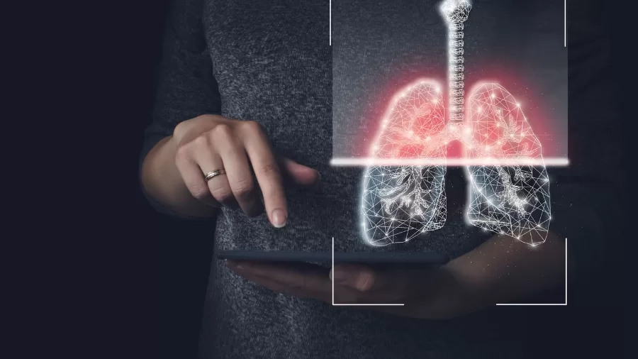
A novel approach to scanning the lungs is changing the way medical practitioners assess and monitor lung health. This approach allows for real-time study of lung function, offering vital insights into therapy effects and detecting diminishing lung function early on. Such developments show potential for improving the care of patients with lung disorders, including those who have undergone lung transplants.
Real-Time Visualisation of Lung Function
Researchers at Newcastle University in the United Kingdom developed a new scanning approach that provides a unique picture of how air moves in and out of the lungs during breathing. This is especially useful for people with asthma, chronic obstructive pulmonary disease (COPD), and lung transplant recipients.
Researchers may safely acquire comprehensive images of lung airflow by employing a specific gas known as perfluoropropane, which is visible on an MRI scanner. The gas that patients inhale and exhale tells which regions of their lungs receive air and which do not.
Professor Pete Thelwall, Project Lead and Professor of Magnetic Resonance Physics at Newcastle University, said: “Our scans show where there is patchy ventilation in patients with lung disease, and show us which parts of the lung improve with treatment. For example, when we scan a patient as they use their asthma medication, we can see how much of their lungs and which parts of their lung are better able to move air in and out with each breath.”
Improved Diagnosis and Treatment
Researchers can use this new scanning approach to identify poorly ventilated parts of the lung and assess overall ventilation efficiency. This feature enables a more accurate assessment of the impact of respiratory disorders.
The team demonstrated the technique’s effectiveness in patients with asthma and COPD, as described in their Radiology report. Researchers have shown that assessing the degree of improvement after delivering medications, such as the widely used inhaler salbutamol, could play an important role in clinical trials for new lung disease therapy.
Advances in Lung Transplant Care
The potential applications of this scanning technique go beyond typical respiratory ailments. A separate study published in JHLT Open looked at lung transplant patients treated at Newcastle upon Tyne Hospitals NHS Foundation Trust. This study demonstrated how the approach could improve the monitoring of lung transplant patients by detecting early changes in lung function.
In cases of chronic rejection—a common condition in which the immune system destroys donor lungs—the scans revealed decreased air movement to the lungs’ periphery, a sign of minor airway damage.
Professor Andrew Fisher, Professor of Respiratory Transplant Medicine at Newcastle Hospitals NHS Foundation Trust and Newcastle University, noted the following possible benefits: “We hope this new type of scan might allow us to see changes in the transplant lungs earlier and before signs of damage are present in the usual blowing tests. This would allow any treatment to be started earlier and help protect the transplanted lungs from further damage.”
Future Prospects
The team feels that this scanning technology will be a significant tool in the management of lung illnesses and transplantation. Its sensitivity and ability to detect early changes in lung function may result in better disease treatment and patient outcomes.
This study, funded by the Medical Research Council and The Rosetrees Trust, marks a significant advancement in respiratory treatment. As technology advances, clinical applications are projected to grow, providing hope to millions of patients worldwide.
This invention, which allows for real-time visualisation of lung function and exact measurements, has the potential to revolutionise the landscape of lung disease diagnosis and treatment.


